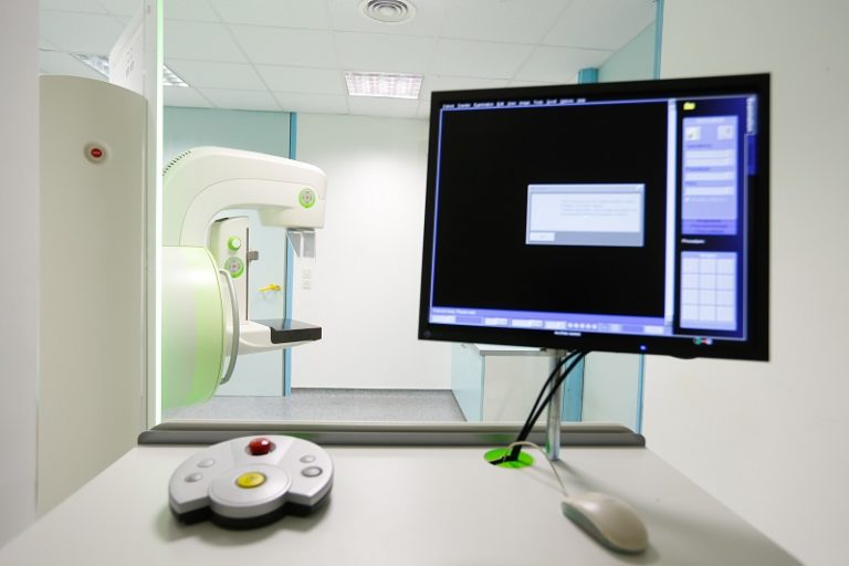
MammoScreen AI: How Artificial Intelligence Supports Mammogram Review

AI Is a Support Tool, Not a Replacement for the Radiologist
Artificial intelligence is increasingly used to support medical imaging, including mammography.
MammoScreen is an FDA-cleared technology designed to assist radiologists as they review mammogram images. It analyzes image patterns and highlights areas that may deserve closer attention.
The radiologist remains responsible for interpreting the exam and issuing the report. AI does not perform the mammogram, speak with the patient or make an independent diagnosis.
Its role is to provide an additional layer of information within the radiologist’s established workflow.
How MammoScreen Evaluates Images
MammoScreen uses a deep-learning model trained on a large collection of mammogram images.
When a new study is processed, the system assesses the images and provides information that can help the radiologist prioritize or re-examine areas of concern.
It can be used with both 2D and 3D mammography images, depending on the clinical setup.
This process happens behind the scenes. Patients do not need additional compression, extra images or a separate appointment simply because AI support is being used.
Your physical mammogram experience remains the same.
Why a Second Analytical Tool May Be Useful
Mammograms contain complex patterns of fatty, fibrous and glandular tissue. Dense tissue can be particularly challenging because it may overlap with or obscure an abnormality.
An AI tool can offer consistent image analysis and draw attention to features that the radiologist should consider alongside the full exam.
AI is not infallible. It can flag benign findings, and it may not highlight every cancer.
Radiologists also consider prior images, breast density, technical quality, symptoms, history and patterns that an algorithm may not fully capture. Human expertise and clinical context remain essential.
What Happens If an Area Needs More Evaluation?
If the radiologist sees an area that is unclear or potentially abnormal, the next step may include additional mammogram views, targeted ultrasound, short-term follow-up or biopsy.
That process is based on the radiologist’s final assessment, not solely on an AI score or marker.
A callback after screening is not a diagnosis of cancer. It means more information is needed to reach a confident conclusion.
Follow the instructions in your results letter and contact the ordering provider if you have questions about the recommended next step.
MammoScreen at Women’s Health and Menopause Center
WHMC includes MammoScreen with mammograms to support the breast-specialized radiologist reviewing each study.
The technology is intended to enhance review while preserving the role of trained clinical professionals.
Learn more on WHMC’s MammoScreen information page.
If you are due for screening, call (248) 932-9223 or request an appointment online.
You can also review WHMC’s 3D mammography services and ask whether additional screening should be considered based on your breast density and individual risk.
Questions Patients Often Ask About AI Mammography
Does AI see my entire medical record? The system is used to analyze mammogram images within a clinical workflow. The radiologist, not the algorithm, brings together the imaging with prior studies and relevant history.
Does an AI marker mean cancer is present? No. A highlighted area is a prompt for review, not a diagnosis.
Can AI replace additional imaging? No. If an area remains unclear, the radiologist may recommend diagnostic views, ultrasound, MRI or biopsy.
Does AI guarantee that a cancer will be found? No screening method or software can provide that guarantee. Patients should still report new breast changes even after a normal mammogram.
The most useful way to think about MammoScreen is as one more analytical input for the radiologist.
It can support consistency and attention, but it works within a broader system that includes high-quality imaging, trained technologists, specialist interpretation, comparison with prior exams and reliable follow-up.
Technology Works Best With Complete Information
Even when AI is part of the review, provide accurate history and make prior images available.
Tell the team about surgery, implants, previous biopsies, current symptoms and changes in family history. Those details give the radiologist context that image analysis alone cannot supply.
Advanced software may support interpretation, but high-quality care still depends on communication among the patient, technologist, radiologist and referring clinician.